Automated Cord Blood Cell Viability and Concentration Measurements Using the Vi‑CELL XR
Introduction

Stem cells, due to their differentiation into mature blood cells, are the key to successful bone marrow transplantation research. More recently, it has been found that umbilical cord blood is also a plentiful and rich source of hematopoietic stem cells.(1) Thus, both bone marrow and cord blood are used in treatment research of numerous cancers, immunological disorders, and certain genetic diseases.
Currently, many facilities provide parents the option to store, or bank, their newborn baby’s cord blood.(2) When cells are banked—usually in liquid nitrogen—two parameters must be accurately assayed. These measurements are cell concentration and percentage of cell viability.(3) These measurements are performed prior to storage and after the thawing process. Cells may decrease in both number and viability, mainly due to the cryopreservative employed in the freezing process (usually DMSO).
The Vi-CELL XR (Figure 1) automates the manual trypan blue vital dye exclusion method for cell viability determinations. In addition, the instrument provides an objective measurement of cellular concentration. As mentioned, these are two critical parameters required in the cord blood cell banking process. The objective of this work was to describe a method for cord blood sample preparation, and develop a cell type, accurate set of instrument parameters, for the Vi-CELL XR.
Materials and Methods
A sample of cord blood was obtained from Baptist Hospital, Miami, FL, by the SAS laboratory at Beckman Coulter. The blood was diluted 1:1 using room-temperature phosphate buffered saline. The standard Ficoll* gradient separation method was used to isolate the mononuclear cells.(4) Isolated cells were washed once in PBS and resuspended in 2 mL of Isoflow* (Isoton* II). A 1:10 dilution of the cell suspension was prepared by placing 100 µL of cells into 900 µL of Isoflow in a standard Vi-CELL™ sample cup. The Vi-CELL XR was used to develop the cord blood cell type. Prior to sample analysis, the Vi-CELL™ concentration control was assayed.Results
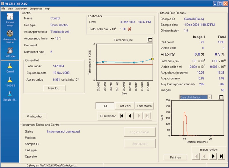
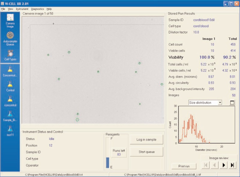
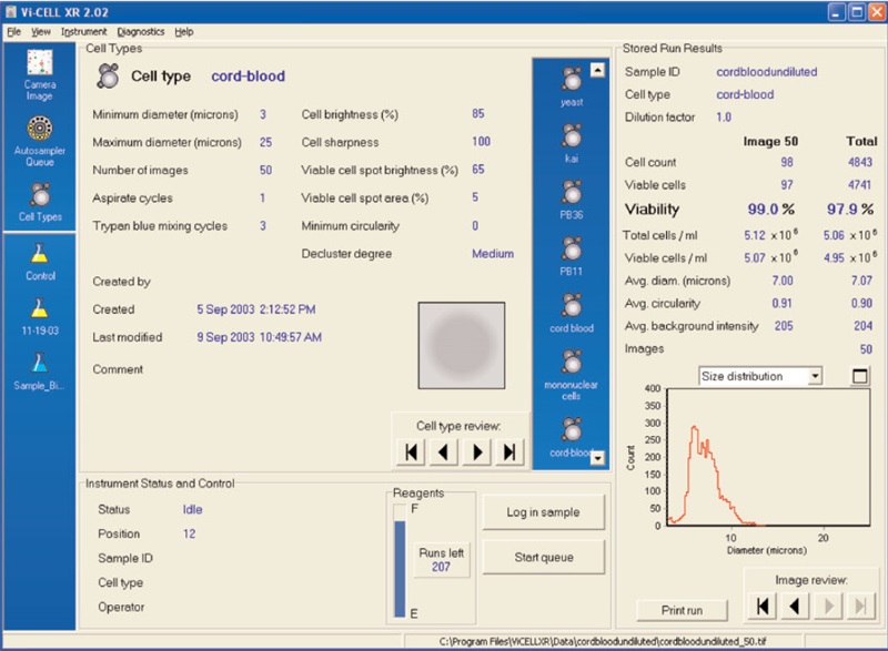
Figure 2 shows the results of the Vi-CELL concentration control. Figure 3 illustrates a cord blood cell image on the Vi-CELL XR. Figure 4 shows the cell type parameters used to analyze cord blood cells. The size range was set from 3 to 25 microns. Cell brightness: 85; cell sharpness: 100; viable cell spot brightness: 65; viable cell spot area: 5.Conclusions
Cord blood stem cell infusions have significant advantages over bone marrow transplants. Viability and concentration of cord blood cells may be assayed using the Vi-CELL XR . The cell type settings for cord blood samples were determined. Size range 3 to 25 microns. Imaging parameters: Cell brightness: 85; cell sharpness: 100; viable cell spot brightness: 65; viable cell spot area: 5.
References
- Kurtzberg, J., Laughlin, M., Graham, M. L., et al. Placental blood as a source of hematopoietic stem cells for transplantation. Blood 90, 4665-4678 (1997).
- American Academy of Pediatrics Work Group on Cord Blood Banking. Cord blood banking for potential future transplantation: subject review. Pediatrics, Vol. 104, No. 11, 116- 118 (1999).
- Fiorino, Susan, Johns Hopkins University, The Sidney Kimmel Comprehensive Cancer Center. Personal Communication.
- Boyum, A. Separation of white blood cells, Nature 204, 793-794 (1964).
Helpful Links
-
세포 계수기(셀 카운터) - 세포 수 계산 및 생존율 계산
-
Vi-CELL BLU 세포 생존도 분석기
- 21 CFR Part 11 Compliance with the Vi-CELL BLU Cell Viability Analyzer
- Vi-CELL BLU Analyzer Integration with Sartorius Ambr® 250 High-Throughput Bioreactor & Ambr® 15 Cell Culture Systems
- Vi-CELL BLU Cell Viability Analyzer Automation Integration
- Automated Bioreactor Sample Delivery to the Vi-CELL BLU Cell Viability Analyzer
- Automated Bioreactor Sample Delivery to the Vi-CELL BLU Cell Viability Analyzer
- Top 3 Reasons to Upgrade Your Lab Instrument
- Virtual Product Demonstration Request
- Webinar Gallery
- Vi-CELL BLU vs. Vi-CELL XR: Comparison of System Specifications
- Vi-CELL BLU Cell Analyzer Features
- Vi-CELL BLU Cell Viability Analyzer Software
- Cell Type Guidelines & Optimization
- Vi-CELL BLU Virtual Demonstration Video Gallery
- Automated vs Manual Cell Counting JP
- Vi-CELL MetaFLEX
- Vi-CELL XR 세포 생존도 분석기
- Multisizer 4e
- Z 시리즈 쿨터 계수기
- Multisizer 3 Coulter Counter
-
Vi-CELL BLU 세포 생존도 분석기
-
자료 센터
- 연구 분야
- 핵심 기술
- Disease Management
-
Biologics Drug Discovery and Development
- Analytical Method Development and Testing
- Biologics Assay Development
- Introduction to Biologics
- Target Discovery, Identification and Selection
- Biologics Drug Discovery
- Preclinical Drug Development
- Flow Cytometry in Cell Therapy
- Biologics Upstream Development
- Biologics Downstream Development
- cGMP Biologics Production
- QA / QC Release Testing
- Disease Research
- Explore Your Industry
-
산업 표준
- USP<643>
- 21 CFR Part 11 - Data Integrity
- Maintaining Data Integrity: Understanding ALCOA Guidance
- European Pharmacopoeia EP 2.2.44
- EU GMP Annex 1
- EU GMP Annex 11
- ISO 14644 cleanroom recovery testing explained
- ISO 21501
- ISO Regulations
- IVD-Regulation (IVDR)
- Regulatory Entities: Separation with Cooperation
- USP 1046
- USP 1047
- USP <788>, <787> and EP2.9.19
- USP <790>
- ISO 11171 Standard
- USP <787>
- 기술 및 방법
- Explore Resources For Your Sample Type
-
기술
- Machine Learning Assisted Analysis for Cytometry Data
- Laser Diffraction for Particle Size Analysis
- Next Generation Sequencing
- Analytical Ultracentrifugation
- Title
- Dynamic Light Scatter
- Dried Reagent Technology
- Accessible Flow
- Acoustic Sample Management
- Elevating Bioprocessing Efficiency with Avanti JXN and Optima XPN Centrifuges
- How the Vi-CELL BLU Analyzer supports You from Data to Insights
- 실험실 자동분주기 기초
- Centrifugation Technology
- Cytobank Training
- SPRI Beads
- Accessible Flow Learning Center
- Flow Cytometry Basics
- Flow Cytometry Data Reproducibility
- Flow Cytometry Panel Design
- The Coulter Principle
- 온라인 문서
- 비디오
- 벡크만쿨터 제품 어플리케이션
-
기술 및 방법
- Cell Counting Methods and Technologies
- Cell Harvesting in Cell Culture: Methods, Technologies, and Process Optimization
- Immunophenotyping
- Cell Maintenance
- Immune Monitoring
- Interaction Quantification & Characterization Using Analytical Ultracentrifugation
- High-Throughput Screening
- Viral Particle Purification Using Centrifugation Methods
- Mass Spectrometry
- spINSIGHTS
- Immunophenotyping
- 연구 분야
